JOURNAL CLUB
Seminal paper:
Studies on Rickettsia-like Micro-organisms in Insects
INTRODUCTION
Hertig M, Wolbach SB. Studies on Rickettsia-Like Micro-Organisms in Insects. J Med Res. 1924 Mar;44(3):329-374.7. PMID: 19972605; PMCID: PMC2041761.
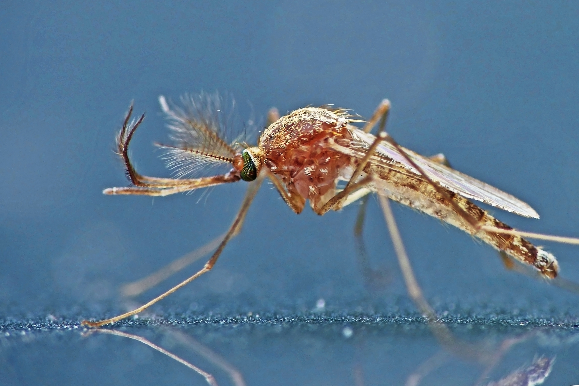
In 1924, entomologist Marshall Hertig and pathologist S. Burt Wolbach published the seminal paper describing the first observations of Wolbachia in the reproductive organs of Culex pipiens. Armed with a binocular dissecting microscope, they provided enhanced staining methods for detecting rickettsia-like microorganisms; criteria for distinguishing the bacteria from cell granules and artefacts; support for the intracellular, hereditary nature of rickettsiae; and morphological observations of various symbiotic relationships between rickettsiae and their arthropod hosts. Throughout the article, they build upon – and dispute – thoeries from other scientists to provide a revised characterization of Rickettsia.
This journal club highlights the iterative process of science presented by Hertig and Wolbach through guided questions, the use of graphic illustrations, and a claim-evidence-reasoning exercise.
Journal Club prepared by Sarah Bordenstein.
GUIDED QUESTIONS
Download the research article.
BACKGROUND RESEARCH
- What is the causative agent of Rocky Mountain spotted fever?
- How is Rocky Mountain spotted fever transmitted?
- What are sheep keds?
- What is meant by “Gram-negative” bacteria?
- Rickettsia and Wolbachia are both members of which taxonomic order?
- What is the common name for Culex pipiens?
INTRODUCTION (pages 329-330)
- Which types of microorganisms were generally described as Rickettsia?
- Based on the authors’ observations, what was the perceived distribution of Rickettsia?
- What was the purpose of this study?
MATERIALS AND TECHNIQUE (pages 331-335)
- Which type of microscope was used?
- Why did the authors prefer smears rather than sections?
- List and describe four criteria used to distinguish rickettsiae from cell granules and other artefacts.
OBSERVATIONS ON VARIOUS RICKETTSIAE
Rickettsia melophagi in the Sheep-Ked Melophagus ovinus (pages 335-340)
- List at least three observations that support the hereditary transmission of Rickettsia-like microorganisms. (page 336)
- What definitive characteristic of rickettsiae was observed by Nöller in 1917 and Jungmann in 1918? (page 336)
- What was meant by the following statement? (page 337)
“We also experienced no untoward results from allowing the keds to feed upon our persons.”
- Describe the dispute between Woodcock and others regarding the nature of rickettsiae. (pages 338-340)
Rickettsia in the Mosquito, Culex pipiens (pages 340-344)
- The authors collected and dissected Culex pipiens mosquitoes from Boston and Minneapolis. What did they observe in all 25 individuals?
- In which organs did they constantly observe this microorganism?
- Describe the observed morphology of this microorganism.
DISCUSSION
- What was the general description of intracellular symbionts? (page 361)
- What was Cowdry’s definition of Rickettsia? (page 363)
- Which characteristics were reportedly missing from Cowdry’s definition? (page 363)
- In conclusion, what was the proposed description of Rickettsia? (page 366)
REFLECTION
- Prior to the internet, how did scientists stay up to date with new discoveries?
- Discuss modern-day tools/techniques that would facilitate the discovery and description of new microbes.
- Isaac Newton famously stated, “If I have seen further it is by standing on the shoulders of Giants.” This phrase has become a metaphor to symbolize scientific progress by building upon the discoveries of others.
-
- List one example from the article in which the authors built upon previous observations.
- List one example from the article in which the authors disputed previous conclusions.
- How are both of these examples important to the advancement of science and our understanding of the natural world?
GRAPHIC ILLUSTRATION
Create a graphic illustration to accompany this research article.
Graphic illustrations capture the interest of readers and concisely summarize a key finding of the research. They often accompany journal publications and news releases to highlight main take-home messages.
Create an illustration for one of the following topics:
#1 – DEVELOPMENTAL CYCLE (Page 343)
The authors describe a tentative cycle of development for the Rickettsia-like microorganisms with respect to their mosquito host (egg – larvae – pupae – adult).
#2 – DESCRIPTION OF RICKETTSIA (Page 366)
The authors propose a revised description of Rickettsia based on cumulative evidence provided in the article.
#3 – TIMELINE OF EVENTS
Create an illustrated timeline to place the results of this paper in context of other scientific discoveries, world events, etc.
TIPS FOR FINDING IMAGES
Google Search for CC Images
- Enter a keyword @ https://www.google.com
- From the main results page, select Images
- From the image results page, select Tools
- Select Usage Rights >> Creative Commons License
Other Resources for Free Images
- Wikimedia Commons: https://commons.wikimedia.org
- Vecteezy: https://www.vecteezy.com
- PhyloPic: https://beta.phylopic.org
- Openverse: https://wordpress.org/openverse
- Flaticon: https://www.flaticon.com
- Bioicons: https://bioicons.com
** If using images from the internet, pay attention to copyright restrictions and include proper citations.
CLAIM – EVIDENCE – REASONING ACTIVITY
Review Figures 4-9 at the end of the article:
What were the observed entities in Giemsa smears of Culex pipiens gonads?
CLAIM:
Write a sentence describing the observations from Culex pipiens smears.
EVIDENCE:
Provide scientific data to support your claim. Use evidence presented on pages 340-343 of the article.
REASONING:
Describe why/how the evidence supports the claim. Use criteria presented on pages 333-335 and throughout the manuscript.

